Discuss the two primary methods of X-ray interactions in diagnostic radiology.
The theory
X-rays interact with matter differently based on the energy of the photon and the atomic number of the material in which the interactions take place. The difference in interactions results in subject contrast in radiology. The two main types of interactions are photoelectric effect and Compton scatter. Subsequent to each interaction, either the photon or the particle will deposit some or all of its energy into the patient tissue which contributes to patient dose. This absorbed dose can lead to adverse health effects.
Photoelectric effect
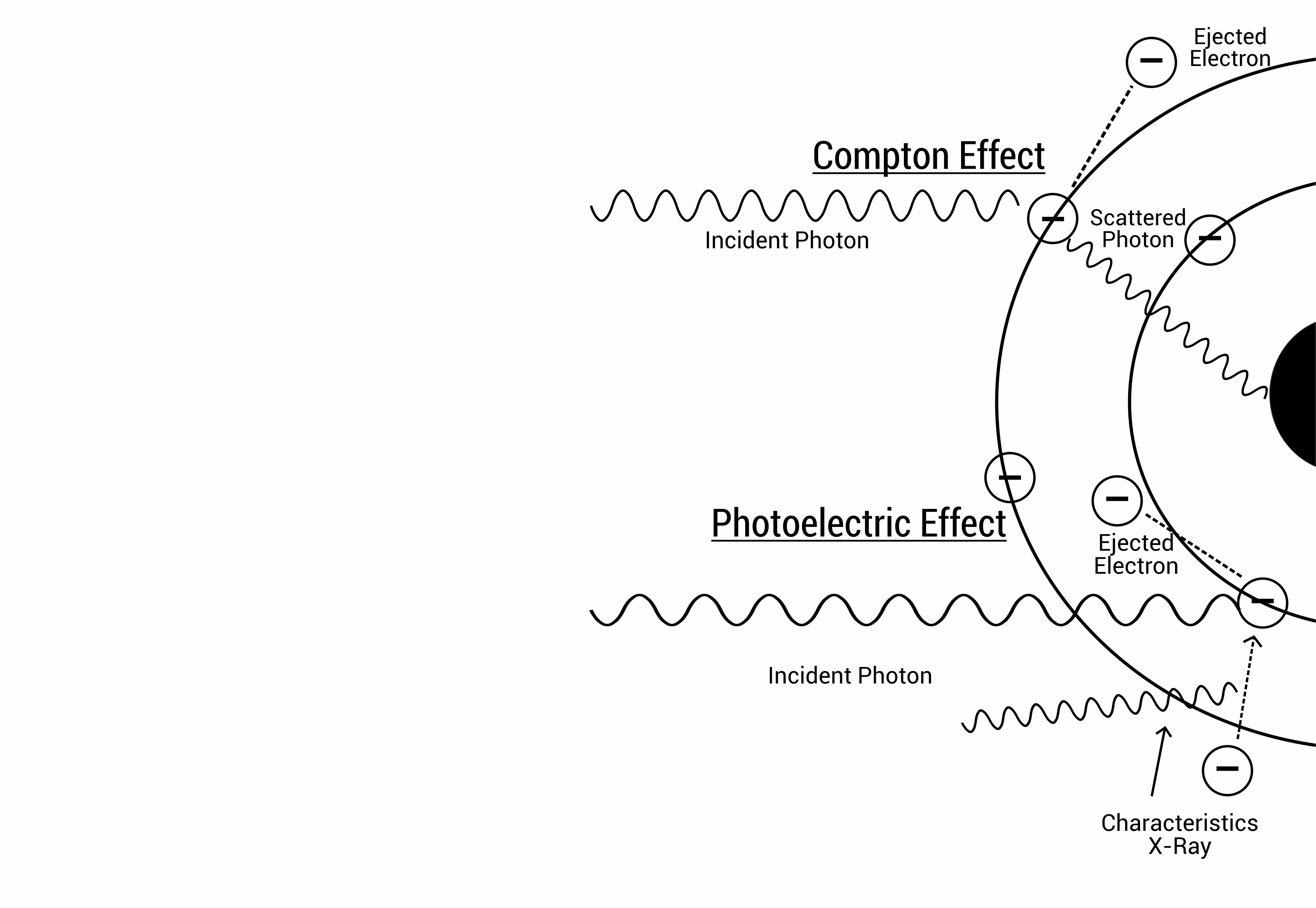
The photoelectric effect is when an incident photon is completely absorbed in the process of ejecting an electron from the atom. The ejected electron escapes with kinetic energy (Ee) which is the difference between the incident photon energy (Ep) and the binding energy of the electron (Eb):
Ee = Ep – Eb
The vacancy left behind is quickly filled by an electron from the outer shell emitting a characteristic X-ray in the process. The characteristic X-ray energy is equal to the energy difference between the higher shell and the vacated shell energies.
The photoelectric effect is most likely to occur at energies just above the binding energy of the K electron. Photons with less energy than this will be insufficient to interact with the K shell but may be sufficient for interactions with the L shell and so on. At energy levels much greater than the binding energy the likelihood for photoelectric effect decreases and different types of interaction is more likely.
The binding energy is also affected by the atomic number of the atom, the greater the atomic number, the greater the binding energy, which means the more likely the photoelectric effect is to occur in the diagnostic energy range. Soft tissue is made up of lower atomic number atoms such as hydrogen, oxygen and carbon while bone is made up of higher atomic number elements such as calcium and phosphorus. Therefore photoelectric absorption is more likely to occur in bone than tissue at conventional diagnostic X-ray energy.
This effect is important in providing subject contrast through differential absorption. The different materials likelihood to fully absorb incident photons differentiates the different structures on radiographs.
In summary, photoelectric effect is most likely to occur in tightly bound electrons at photon energies just greater than the electron binding energy. The chance of photoelectric absorption decreases with increasing photon energy by approximately 1/E3. The likelihood of photoelectric absorption increases with the atomic number by approximately Z3.
Compton effect
Compton effect is when an incident photon is deflected following interaction with an electron. In the process, a portion of the incident photon energy is transferred to the electron, ejecting it from its shell, and the remainder of the energy is carried by the scattered photon at an angle ϴ to its original direction.
The scattered photon becomes a secondary radiation source which can go on and penetrate without any further interaction, or it can further interact undergoing photoelectric absorption or Compton scattering.
The Compton effect is very prominent in the diagnostic energy range. It decreases with increased photon energy. However, the Compton rate decreases much less rapidly than photoelectric absorption rate with increased photon energy. This means as the photon energy increases the relative chance of Compton scatters becomes greater within the patient tissue.
The effect is almost independent of atomic number. Any loosely bound electron (low binding energy) can be subjected to this effect. This means electrons in lower energy shells in low Z number materials and higher energy shell electrons in high Z number materials. Increasing the number of electrons (electron density) enhances the likelihood that a photon would interact with an electron. Therefore the Compton effect is proportional to the electron density of the material.
The secondary photon can be scattered in any direction. However, there is an increased probability for certain angles based on the incident photon energy. Increasing the photon energy will improve the chances for forward scatter. In the diagnostic energy range, however, there is a significant probability for side and back scatter which contributes to increased patient and staff dose.
In summary, Compton effect becomes the more likely interaction type with an increase in photon energy in the diagnostic imaging range. Increasing the electron density of the material enhances the chance of Compton effect. Compton scattering degrades image quality and increases patient dose through secondary photons.

Figure 1 – In the diagnostic radiology energy range, the two most likely effects are Photoelectric absorption and Compton Scattering.
The question
Discuss the two primary methods of X-ray interactions in diagnostic radiology.
In this question it is important to address three things:
State and define the two effects.
Relate the two effects to radiology.
Give a relationship with incident photon energy.
In the first part of this question, the student should define the photoelectric effect and the Compton effect. Following that the student should comment on the how these two interaction methods relate to radiology. Lastly, the student should discuss the effect of photon energy and material on the likelihood of interaction.
The sentence structure should be, “The photoelectric effect is ____. The difference in absorption rates in different tissues leads to image contrast. The effect ____ with increased photon energy and ____ with an increased atomic number. The Compton effect is ____. The scattered photons lead to increased patient dose and increased image noise. The effect _____ with increased photon energy and ______ with an increased atomic number.”
Sample answer
Discuss the two primary methods of X-ray interactions in diagnostic radiology (6 marks)
The photoelectric effect is when the incident photon is fully absorbed by an inner shell electron, ejecting the electron in the process. This leaves a vacancy in the inner shell which is filled by electrons in higher energy states. The difference in photoelectric absorption rates in different tissues leads to image contrast. However, too much photo electric absorption will result in a loss of signal. The photoelectric effect rate decreases with increased photon energy and increases with an increased atomic number. The Compton effect is when an incident photon interacts with a loosely bound electron, ejecting the electron and deflecting with less energy in the process. The secondary scattered photons lead to increased patient dose and increased image noise. The effect decreases with increased photon energy, however much less rapidly than the decrease in photoelectric effect and increases with increased atomic number due to the increased electron density.
Did you find this useful or would you answer this question any differently? Comment below.
One thought on “Exam Question: X-Ray Interactions with Matter”
Comments are closed.